
 فارسی
فارسیGuide
Microtome sectioning problems
Microtome sectioning problems
When studying tissue under a Microscope, the thickness of the tissue should be large enough (in microns) to allow light pass through to be studied by Microscope, otherwise no image will be seen in the Microscope. Therefore, to study the tissues, they must be cut with special tool very thin(in microns) so that they can be seen under a Microscope.
Sample cutting with Paraffin mold is done by a device called Microtome in 5 to 10 microns thickness.
For this reason, the cutting operation is more sensitive than the previous steps and should be done slowly and carefully.
The sharpness of Microtome blades is directly related to the quality of the sections. In Microscopic studies, there should not be any scratches on length or any non-harmonized shape on the sections.
Permanent blades should be sharpened periodically with special tools. The edge of the blades should not be visible under a microscope.
The following are some common problems in Microtomy of the tissues and the causes of these problems.
Some common problems:
-
Very thin cut
Wrong Microtome settings, applying hot breath to cold blocks to accelerate sectioning , selecting the first section from the .ribbon cut, sectioning at very high speeds, inappropriate preparation, lack of Microtome calibration
 figure 1: very thin section
2. Knife lines
damaged knife or blade, inappropriate tissue preparation process, presence of hard materials such as calcium in the block, presence of impurities in paraffin, deposition of buffer salts in the sample
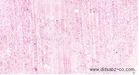
figure 2:existence of knife lines
-
Rupture
Poor preparation process (incomplete dehydration or incomplete clarification and immersion), use of very hot water bath, rough spreading of tissue
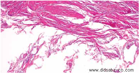
figure 3: tissue rupture
-
Fine cracks
Excessive tissue preparation, very cold block, very fast cut, poor clamping mechanism, improper angle
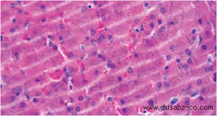
figure 4: fine cracks in the section
-
Twisting
Poor float technique, poor fixation, hot block, very thin cut, very hot water bath
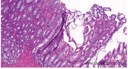
figure 5:existence of torsion in the section
-
High compression
poor preparation process, hot block, very fast cutting, poor paraffin quality, slow cutting edge, very high angle
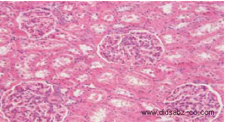
figure 6:high compression of the sample
-
Existence of Bubbles
Existence of Bubbles in Immersion Bath, Poor Immersion Techniques
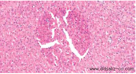
figure 7: existence of bubble in the section
-
Excessive flattening
very high bath temperature, prolonged exposure to water, poor fixation (Solution not removed completely)
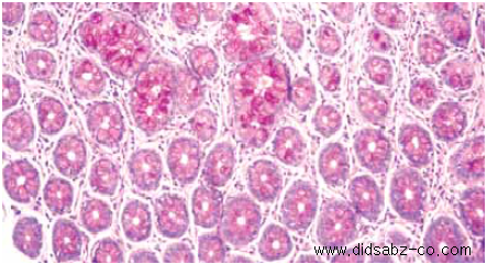
figure 8: excessive tissue flattening
-
Contamination
Dirty slides, contaminated immersion bath, labeling pencil effects
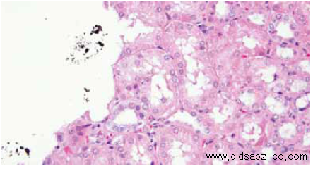
figure 9: existence of contamination