Education
H&E tissue staining
H&E tissue staining
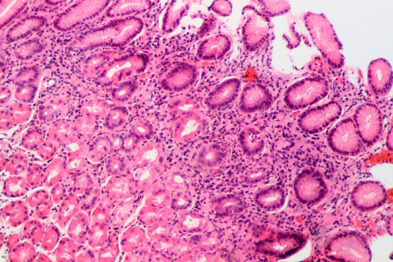
general H&E staining is a method that is commonly used in most pathology laboratories. It also known as Hematoxylin-eosin. This name is actually derived from the name of the material used to perform this method. In this method ,the nucleus and cytoplasm are distinguished and shown with two different colors.
The cell nucleus turns to purple and the cytoplasm turns to pink.
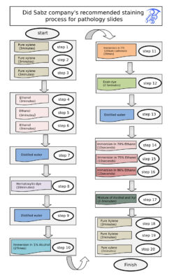
In the following, you can see the staining process recommended by Did sabz company for pathology slides as follow
The recommended staining program is as follows:
Stage1 : pure xylene (2 minute)
Stage2 : pure xylene(2 minute)
Stage 3 : ethanol (3 minute)
Stage 4: ethanol (3 minute)
Stage 5 :ethanol (3 minute)
Stage 6: ethanol ( 3 minute)
Stage 7: distilled water
Stage 8 : hematoxylin dye ( 10 minute)
Stage 9: distilled water
Stage 10: agitation in 1% alcohol acid ( 2 times)
Stage 11: agitation in 1% lithium carbonate (2 times)
Stage 12: eosin dye ( 2-5 minute)
Stage 13: distilled water
Stage 14: agitation in 70% ethanol (10 seconds)
Stage 15: agitation in 75% ethanol (10 seconds)
Stage 16: agitation in 96% ethanol (10 seconds)
Stage 17:Mix alcohol and xylene (2 to 3 minutes)
Stage 18: Pure Xylene (2 minutes)
Stage 19: Pure Xylene (2 minutes)
Stage 20: Pure Xylene (2 minutes)
Did sabz company’s recommended staining process

 فارسی
فارسی

