
 فارسی
فارسیEducation
processing the bone tissue and decalcification
Processing bone tissue and decalcification :
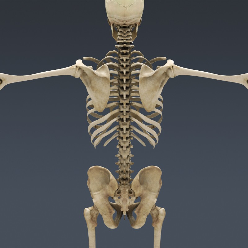
Samples such as bones and teeth, generally the tissues with calcareous deposits should be decalcified before fixation to be prepared for later stages.
Decalcification means to release the minerals (calcium) from bone tissue and involves the following steps:
- Sample preparation
- Tissue Fixation
- Decalcification
- Neutralize and rinse with water
Decalcified bone is used to study the bone marrow to diagnose tumors and infections and other purposes, a notable area of the tissue sample is calcified then, it is impossible to achieve the desired tissue sample without initial declassification.
The decalcification solution should hold the following properties:
- It should Release calcium totally from the tissue
- It should not damage the main tissue
- It should not disturb the staining process
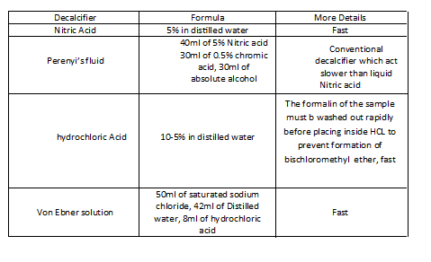
table 1: Decalcification solutions used in the laboratory
Bone is made up of osteocytes, collagen matrices, and crystalline hydroxyapatite , these crystals dissolve during the Decalcification process and if done correctly, the bone tissue sample should be physically Similar fibrosis connective tissue after Decalcification process from density point of view.
Note:
The time required for decalcification depends on the type of sample, If the sample is bone marrow, the time required is one hour Or if it is a femoral specimen, the required time is one month. Increasing the temperature speeds up the process but may damage the tissue so be careful.
Note:
In addition to bone, other tissues, such as the walls of blood vessels or the kidneys and lungs, can also be calcified and require decalcification process before tissue preparation process.
it can be soaked in 10% stick acid To soften semi-hard bone tissue And in order to soften very hard bone tissue, it can be soaked in 10% nitric acid.
Note:
It is better to make a 2.5 mm section of tissue.
Note:
The nature of the sample received is important And should be considered as the amount of Spongy and compact bone is important to determine the time required for declassification and the process.
Determining the end point of the decalcification process
To achieve high quality results, it is important to determine the point at which all the calcium in the tissue is removed is important Excessive continuation of the process causes tissue damage and the reason is the use of strong acids And incomplete process causes inability in the sectioning.
The best method, especially for large specimens such as the femur, is to use X-ray analysis. This analysis detects even the smallest calcium available. A chemical test to determine the presence of calcium is to use ammonium oxalate If calcium oxalate is formed The declassification process should continue. Physical testing including bending and manipulation of the specimen can .also be used, However, the results of this test are not reliable and can also cause damage to the sample
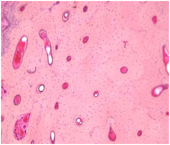
Figure 1: Cross section of long bone decalcified and stained by H&E method
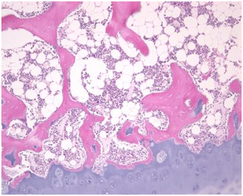
Figure 2: Declassified cross section of spongy bone (pink) and hyaline cartilage ( blue)
Bone powder
But if a metabolic bone insufficiency is examined And it needs to differentiate mineralized bone from osteoids Or morphological measurements of bone required then It is necessary to preserve the mineral part of the bone. And sectioning from Calcified bone Because mineral bones are a very hard tissues and There are limited ways to cut them. It can be spread directly on a silicone plate after fixation And powdered by rough surfaces and powdered thin cross section Prepared as in Figure 6. Another solution is to soak bone samples by impregnating them with acrylic or epoxy resins. so that after polymerization, they have the same hardness as mineral bone ,Then it can prepare slices of the impregnated sample by heavy Microtomes with tungsten-carbide or diamond blades as shown in Figure 3
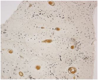
Figure 3: A powder-coated section without staining of compressed bone. These bony layers contain spaces that contain osteocytes. In dried powder sections as shown, they are marked by black structures
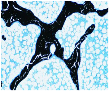
Figure 4: A sample without decalcification of spongy bone. The bone is fixed in formalin and impregnated with epoxy resin. The calcified bone is marked in black and the steroids are painted blue on the trabecular bone. We see that despite the support of epoxy resin, the calcification matrix has cracked when preparing the cut.
I’m juggling about 10 things right now so I don’t have that much time to play around learning how to make a website. What are good resources to jump-start implementing javascript, php, mySQL, etc?.