
 فارسی
فارسیEducation
Tissue staining
Tissue staining stage :
this step includes 2 types of general and specific methods . the general methods is : Hematoxylin-eosin method , with this method the nucleus turns to purple and cytoplasm turns to pink.
The specific method is that each cell component takes a specific color, which includes staining of frozen sections , connective tissue , neural system , staining for some cytoplasmic materials , staining for enzymes , staining for micro organisms and cytological staining.
Solutions used in routine staining include : Ethanol, xylene, Hematoxylin , Eosin , Hydrochloric Acid, lithium Carbonate, Distilled water, Acetic Acid.
Note:
0.5 cc of Acetic Acid should be added to every 100 cc of Hematoxyline before use.
Note:
Place the sections in the oven at temperature of 120◦ C for 15-20 minutes before staining , letting them stick completely to the slides.
Procedure:
- pure xylene (1-2 minutes)
- pure xylene (1-2 minutes)
- pure xylene (1-2 minutes)
- ethanol 96% (2-3 minutes)
- ethanol 80% (2-3 minutes)
- ethanol 70% (5 minutes)
Rinsed with distilled water( temperature about 36 degree)
7. Hematoxylins : 5 to 10 minutes, red
Rinsed with distilled water
8. 1% alcohol acid (prepare 70% alcohol and pour 1 cc of concentrated hydrochloric acid for every 100 cc of alcohol)
9. 1% saturated lithium carbonate: several dips (per 100cc of distilled water, one gram of lithium carbonate powder, blue)
10. Eosin (4 to 6 minutes)
Rinsed with distilled water
11. 70% alcohol (1-2 minutes)120
12. alcohol 80% (1-2 minutes)
13. alcohol 96% (1-2 minutes)
14. alcohol 96% (1-2 minutes)
15. mixture of alcohol and xylene (1 volume alcohol 96%, 3 volume xylene)
16. pure xylene (1-2 minutes)
17.pure xylene (1-2 minutes)
18.pure xylene (1-2 minutes)
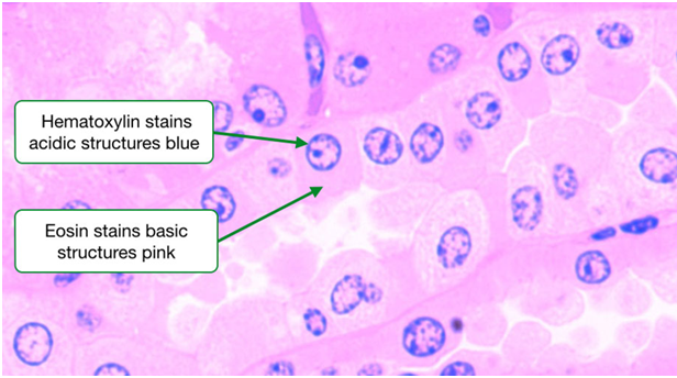
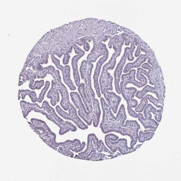
Figure1: an example of Kidney Tissue stained by H&E method
Note:
This method of staining strongly highlight the nucleus of the cells that have turned to blue color, & also their morphology & location.
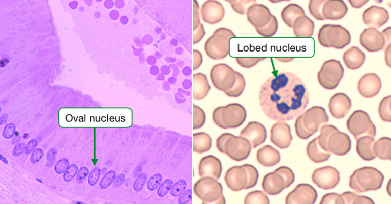
Figure2: detection of nucleus , location & morphology of cells by H&E method
It is also possible to observe the density of the cells in a specific area & the divided cells by H&E staining under a microscope.
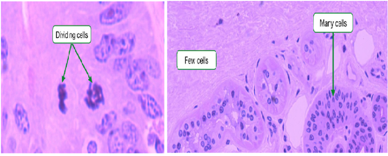
Figure 3: identification of divided cells & cells density by H&E method.
Using this method it is possible to size the cells & observe their structure and that is because all tissues contain blood vessels & this leads to the possibility of comparing the size of the tissue cells.
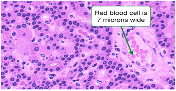
Figure 4: estimating the size of the cells by H&E staining method
this is nicely said.